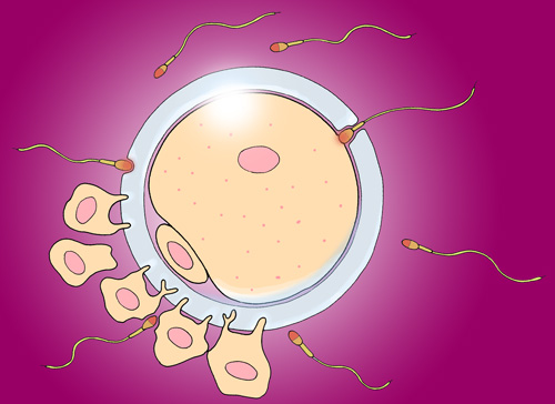
Day 1 - Development of the new individual begins when a spermatozoon penetrates the jelly-like zona pellucida and contacts the plasma membrane of the secondary oocyte. Note the smaller first polar body alongside the secondary oocyte within the zona pellucida. This was formed during the first meiotic division. Several follicular cells are shown still attached to the outer surface of the zona pellucida. After the arrival of the fertilising sperm, no further spermatozoa are allowed to enter.

Day 1 continued - In response to the fertilising sperm, the oocyte completes its second meiotic division. The resulting cell division is unequal - the larger daughter cell becomes an ovum, and the other much smaller daughter cell becomes the second polar body. While the oocyte is dividing, the first polar body may also divide. The head of the spermatozoon enters the cytoplasm of the oocyte and swells, forming the male pronucleus. (The mid-piece and tail of the spermatozoon do not enter the ovum.)
Day 1 continued - After completing its second meiotic division, the nucleus of the larger ovum becomes the female pronucleus. The male pronucleus swells, and the two pronuclei approach each other and merge. This establishes the diploid genome and also the genetic sex of the new individual. The fertilised ovum is called a zygote.
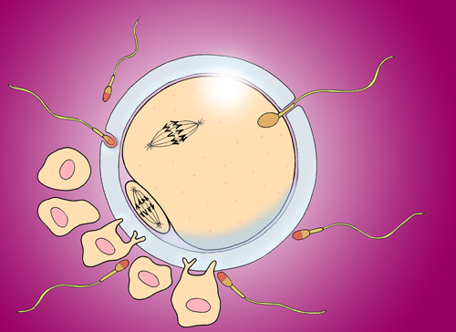
Day 1 continued - The nuclear DNA is replicated and after several hours the zygote begins its first mitotic cell division. This illustration shows metaphase, with the chromosomal pairs arranged at the equator of the mitotic spindle ready to be separated and moved to opposite ends of the spindle.
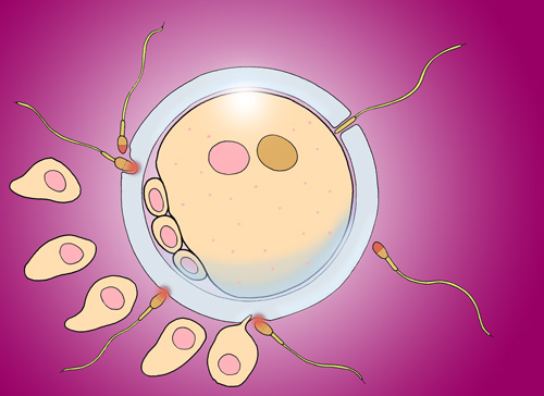
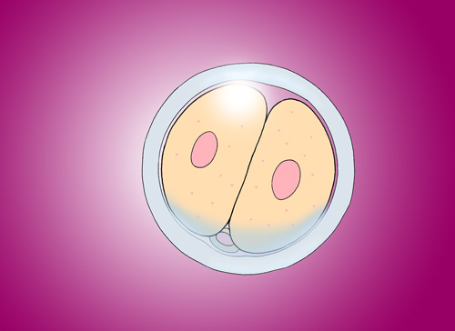 Day 2 - The two-cell stage. The cytoplasm of the original zygote has been subdivided to form two smaller cells - there is little synthesis of new cytoplasmic material at this time. This also applies to subsequent cell divisions during the first few days, so they are often called ‘cleavage divisions’. The cells remain enclosed by the zona pellucida. The polar bodies are less prominent, and probably degenerate.
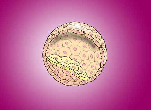
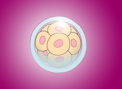 Days 2, 3, 4 - As cleavage continues, a closely-packed ball of cells is produced - the morula. Each one of these cells is still very high in developmental potential, and could each give rise to a new individual, although usually they will co-operate in the development of just one baby.
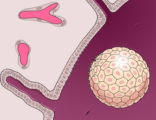
Day 5 - The morula drifts along the fallopian tube as cleavage continues. When it reaches the uterus, the zona pellucida is softened and dissolved by an enzyme in the uterine fluid and releases the morula.
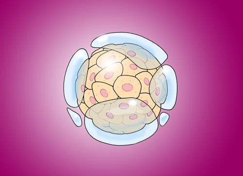
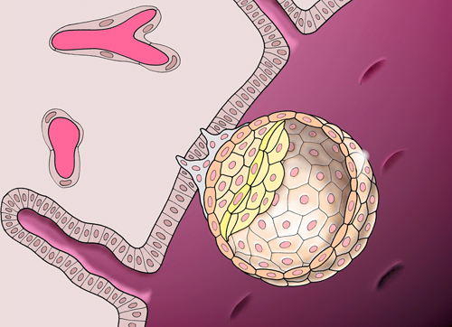
 Day 6 - Fluid is drawn into the centre of the morula, creating a hollow blastocyst.
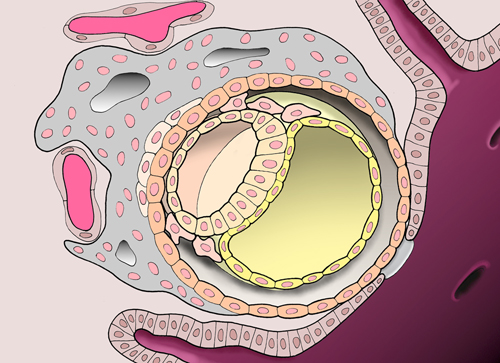
Day 6 continued - Within the hollow, fluid-filled blastocyst there is a special groups of cells called the inner cell mass. The cells forming the outer surface of the blastocyst are called trophoblast cells.
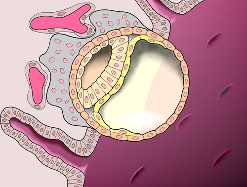
Day 7 - the blastocyst approaches the lining of the uterus - the endometrium. This has prepared for such a possibility by thickening, becoming more glandular, and developing a rich blood supply.
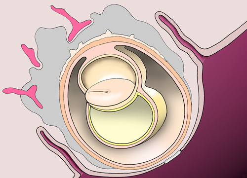
Day 7 continued - The blastocyst begins to implant, a process that will take several days. Implantation brings the outer cells of the blastocyst into direct contact with the maternal cells of the endometrium. The conceptus is genetically different from the mother since half its chromosomes have come from the father. Usually, genetically different cells would be rejected by the mother’s immune system, but this does not occur during normal pregnancy.
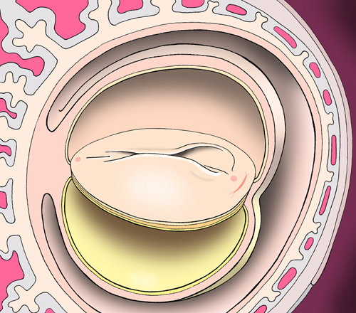
Day 8 - During implantation, the trophoblast thickens and forms two layers - an outer syncytiotrophoblast (grey) and an inner cytotrophoblast. Meanwhile, the inner cell mass becomes organised into a two-layered plate of cells called the embryonic disc, with the amniotic cavity above and the yolk sac below. The two layers are called ectoderm and endoderm.
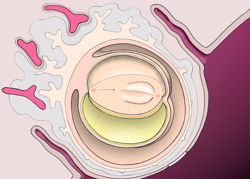
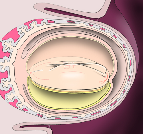
Day 9 - The conceptus implants itself completely within the endometrium. The enlarging syncytiotrophoblast develops fluid-filled lacunae within it and comes into contact with the maternal blood vessels. Extra-embryonic mesodermal cells derived from the cytotrophoblast begin to form a layer around the external surfaces of the amnion and yolk sac.
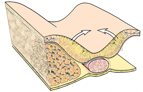
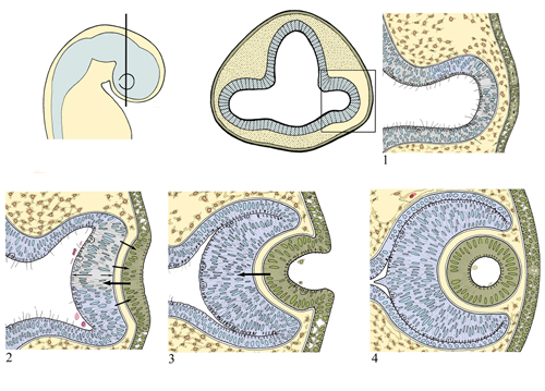
Day 12 - The embryonic disc will give rise to the baby itself. The first sign of the midline axis of the baby is the formation of the primitive streak in the ectodermal layer facing the amniotic cavity.
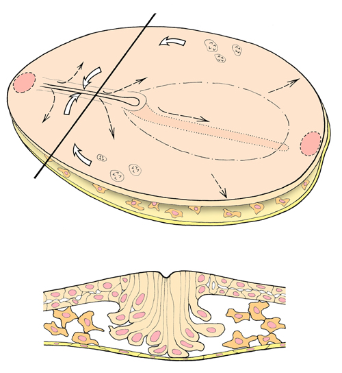
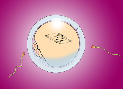
Day 17 - In this diagram, part of the embryonic disc has been removed to show the primitive streak in cross-section. Ectodermal cells migrate towards the primitive streak and then tuck inwards to form a new layer of cells in between the ectoderm and endoderm. The new layer is called the mesoderm. These three layers of cells - ectoderm, mesoderm, and endoderm - then give rise to all parts of the baby's body by a process called morphogenesis.
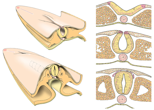
Day 17 continued - The primitive streak is situated in the midline towards the future tail end of the embryo. Further towards the future head end, the ectoderm thickens to form the neural plate. This begins to fold, initially forming a groove and then closing over to form the neural tube. The neural tube will give rise to the brain and spinal cord.
Day 19 - Closure of the neural tube begins in the future hindbrain region and then continues for several more days, extending both forwards and tailwards until closure is complete.
Day 20 - Clusters of mesodermal cells produce pairs of somites alongside the developing neural tube. The number of somites increases steadily as neural closure continues.
Day 21 - In the future brain region, the neural folds enlarge as they close and begin to overshadow the part of the embryonic disc that lies further forwards. Within the mesoderm of this part the heart begins to develop.
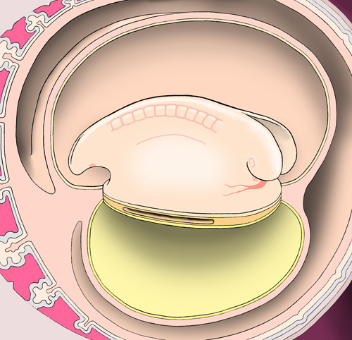
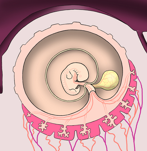
Day 23 - As the heart develops, blood vessels form in the wall the yolk sac, the embryo itself, and in the stalk that connects the embryo to the trophoblast. Blood cells are also formed in the yolk sac wall. On day 23 after fertilisation, the heart begins to beat and the circulation of blood is established. The eyes and ears are developing.
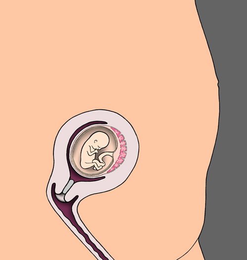
Day 25 - As the embryo develops, the amniotic sac enlarges and gradually the yolk sac begins to diminish in relative size. The surrounding trophoblastic tissue develops finger-like processes which extend outwards into the endometrium. These extensions are best-developed across the most deeply-implanted part of the conceptus, and will contribute to formation of the placenta.
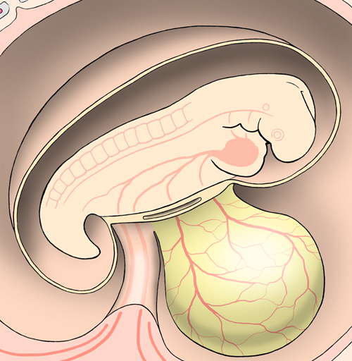
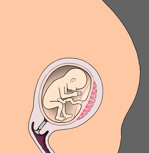
Day 27 - Limb buds begin to form alongside the neural tube and somites. The developing eyes are visible, and the beating heart is now tucked ventrally in the future thoracic region.
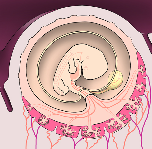
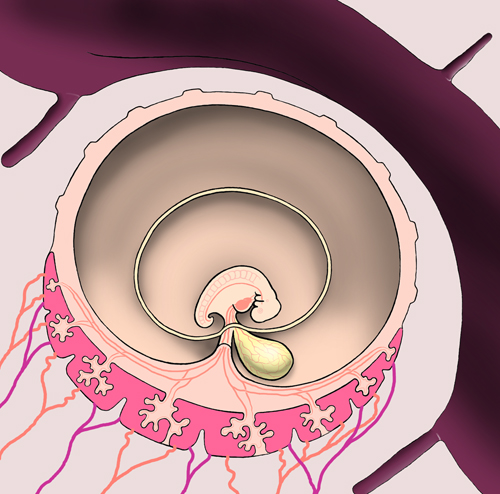
Day 32 - The fingers and toes are becoming apparent, and the external ear is visible on the side of the neck region. The connection between the embryo and surrounding trophoblast is becoming the umbilical cord, and passing through this is a narrow duct connecting the yolk sac with the developing digestive tract in the embryo.
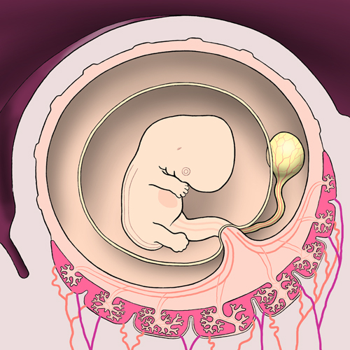
Day 40 - The fingers are separating as the cells forming the webs between them die, a process called apoptosis. The toes are less advanced at this stage.
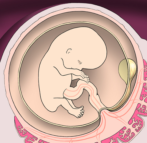
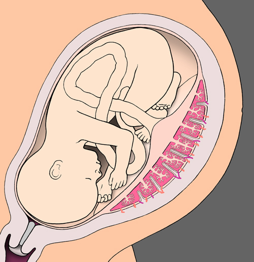
Day 56 - By the end of the second month, all the different parts of the new individual have formed. Morphogenesis is complete. However, the embryo is only about 3 cms long from the top of its head to its rump. Most of the body organs and systems are only partially functional.
Three months - The fetal period begins. It is a time of rapid growth in size and weight, the further maturation of function in organs and body systems, and the rehearsal of increasingly complex activities that must be perfected before birth - breathing movements, swallowing, production of urine, and digestion, for example.
Five months - The uterus and placenta continue to enlarge to accommodate the growing fetus and meet its increasing needs.
Eight months - Towards the end of pregnancy, space is at a premium, and the placenta is finding it increasingly difficult to meet the needs of the fetus.

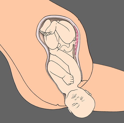
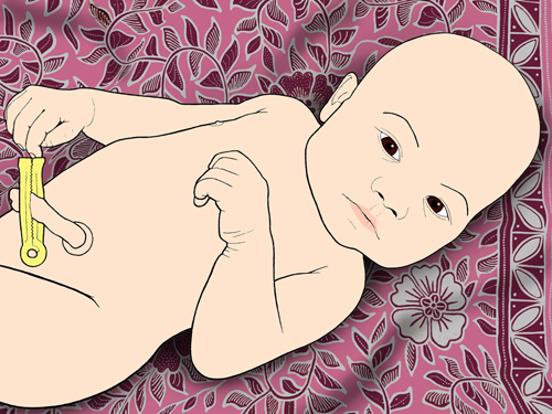
Nine months - The process of birth takes place. A difficult transition must be achieved with the baby’s systems rapidly taking over many of the responsibilities that were met previously by the placenta and mother.
|