


| | | During the embryonic period, all the major organs and systems of the body are formed - in miniature. Morphogenesis is the name given to the process by which bodily form is established (morphos means ‘form’ and genesis means ‘origin’). 
| The key embryonic processes that contribute to morphogenesis are: | cell division | | changes in cell shape | | folding of cell layers | | cell migration | | cell death (apoptosis) | | cell-to-cell communication | | differentiation |
Embryonic discThe cells of the inner cell mass become organised into two hollow spheres in contact with each other and attached to the inside of the trophoblastic shell. One of the inner spheres encloses the amniotic cavity, and the other encloses the yolk sac cavity. Where the two inner spheres contact each other there is a two-layered embryonic disc.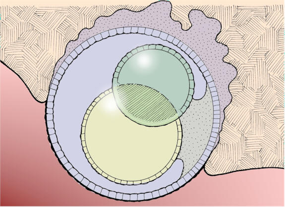
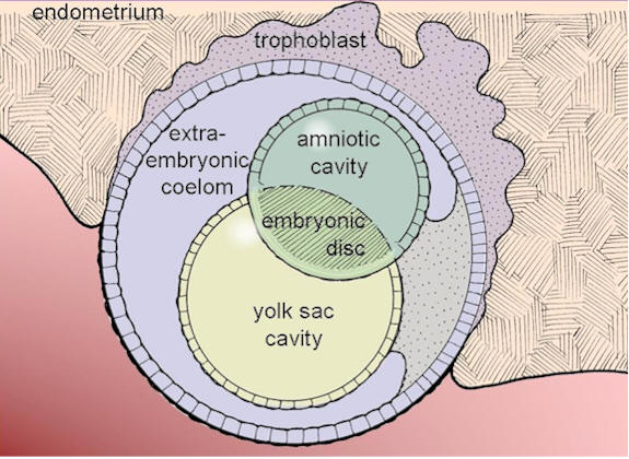
Embryonic disc situated between the amniotic and yolk sac cavities
Cell divisionCell division continues throughout the prenatal period. Most of these divisions are mitotic (meiotic divisions occur only in germ line cells destined to form gametes). Dividing cells - and hence the embryo - are very sensitive to a variety of chemical and physical agents.Changes in cell shape Shape changes have a central role in morphogenesis. They are produced by the co-ordinated activity of the cytoskeleton. The cytoskeleton consists of microtubules and different types of microfilaments. Microtubules assist in elongation of cells and movement of structures such as the nucleus and chromosomes. Microfilaments can draw structures closer together.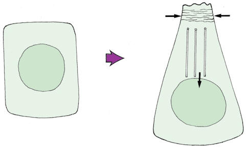
Microfilaments and microtubules
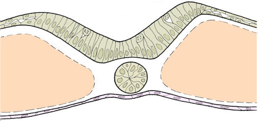
Folding of cell layersWhen shape changes occur within cells forming an epithelium, the epithelium will buckle and fold. This occurs frequently during morphogenesis, and can give rise to tubular and spherical forms. Examples are seen in the formation of the central nervous system and the lens of the eye. 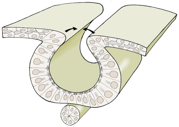 Folding of an epithelium
Cell migrationDuring morphogenesis, many cells migrate within the embryo. Mesoderm cells move and aggregate to form structures such as the somites. Neural crest cells arise from the ectoderm alongside the developing neural tissue and migrate amongst the mesodermal cells.
Cell death It is perhaps surprising to discover that some embryonic cells have to die for normal development to occur. For example, normal development of the fingers and toes depends on death of the cells forming webs between them. The cells die without initiating an inflammatory response - this process of closing down is called apoptosis.
Cell-to-cell communicationEmbryonic cells ‘talk’ to each other - cell-to-cell interactions co-ordinate development and influence gene-selection. Some regions of the embryo are induced to develop in the correct way by chemical signals from neighbouring tissues, eg: the lens of the eye. Induction is just one aspect of a continuous network of interaction linking cells.
DifferentiationAll the cells of the embryo are derived from the zygote, and they have the same genetic information in their nuclei. However, during development differences arise between cells. Different selections of genes are made in each cell type. As cells become more differentiated, they lose developmental potential and become more committed to a particular fate.
Embryonic discInitially, the embryonic disc has two layers - ectoderm and endoderm. Then a third layer called the mesoderm is formed by invagination of ectodermal cells through the primitive streak. The three layers are referred to as the germ layers - they give rise to all the organs and systems of the baby’s body.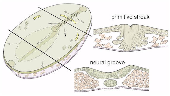
Sections through the embryonic disc in the region
of the primitive streak and neural plate 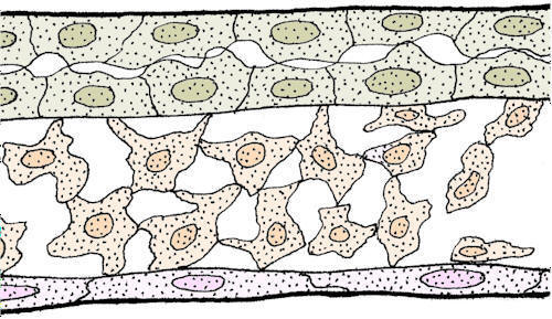
Germ layers 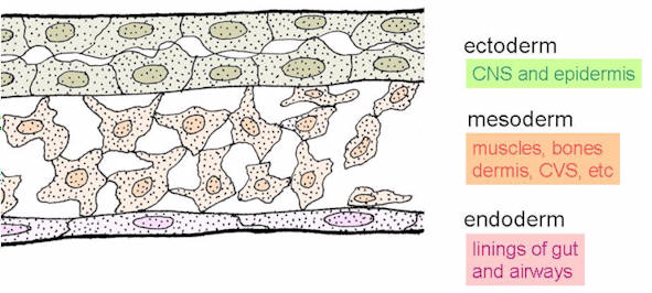
Contributions by germ layers to parts of the body Some of the mesodermal cells aggregate to form the midline notochord, a precursor of the axial skeleton. The ectoderm over the notochord thickens to form the neural plate. The embryonic disc remains two-layered in the region of the oral and cloacal membranes.
Folding of neural plateThe neural plate begins to fold, forming the neural groove. Closure to form the neural tube begins in the future midbrain region, and then extends cranially and caudally. Neural crest cells migrate away from the site of closure and later take numerous roles within the body, forming peripheral neurons, Schwann cells, melanocytes, and head and neck structures.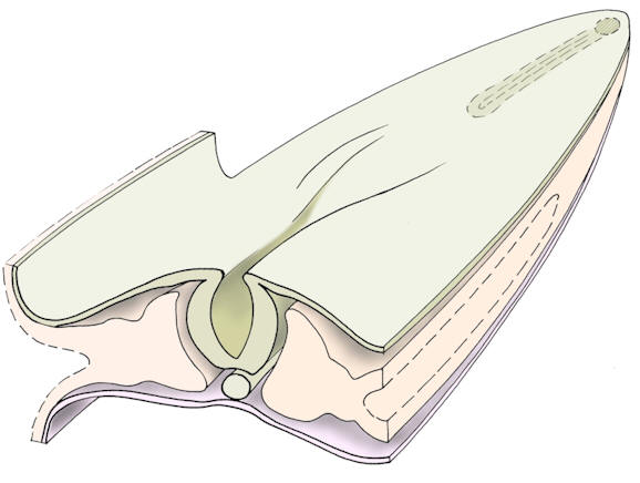
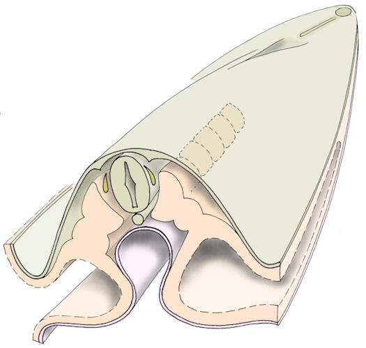
Folding and closure of the neural plate to form the neural tube 
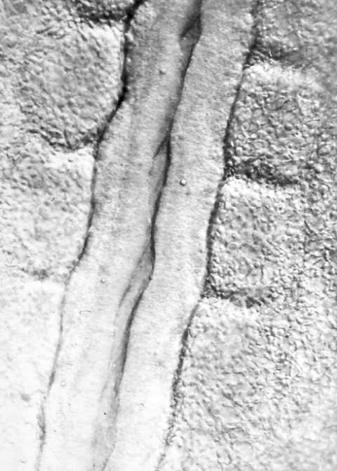
Developing neural tube in a living chick embryo 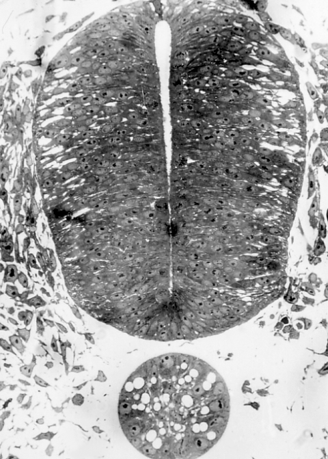
Section through chick neural tube

Sections through the neural plate as it closes to form the neural tube
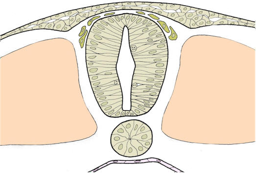
Formation of somitesMesodermal cells form up alongside the neural tube and notochord to form segmental somites. Later, the somites will contribute to skeletal elements, muscles, and dermis. This segmental origin is apparent in the pattern of spinal nerves, vertebrae, ribs, nerve supplies, and dermatomes in the completed body.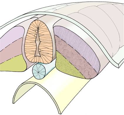
The three main zones of somites - sclerotome (yellow),
myotome (brown), and dermatome (purple) The sclerotome will go on to form skeletal elements such as the vertebral column and ribs, the myotome will form muscles, and the dermatome will form the dermis of the skin. |
|
|