


| | | This is about the first week of development, from the time that the sperm makes contact with the oocyte (fertilisation or conception), to the time that the blastocyst begins to bury itself into the mother's endometrium (implantation). 
| Gametes 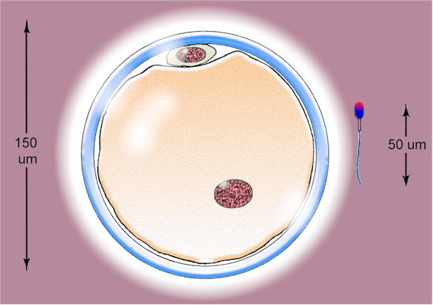 A comparison of the sizes of the two gametes
the larger oocyte is on the left, and the sperm on the right The gametes are haploid - they contain only half the normal number of chromosomes. This is a result of meiosis, a special kind of cell-division. At fertilisation, 23 chromosomes are contributed by the oocyte and 23 by the sperm. Fertilisation restores the diploid number – 46 chromosomes.For each chromosome inherited from Mum, there is usually a matching chromosome from Dad. Chromosomes in a matching pair are called homologous chromosomes. Therefore, each cell has two copies of most genes, one version from Mum and one from Dad.Sex chromosomesThe exception to this rule occurs in males – the sex chromosomes in the male (X and Y) are different in size and in the genes they carry, so that some genes are not duplicated. In the female there are two X chromosomes – XX – so the genes are duplicated.AllelesThe duplicate copies of genes on homologous chromosomes are mostly identical, but may differ. For example, the genes and other factors that control eye colour provided by the maternal chromosomes may be different from those coming from Dad. When we have two different forms of a gene for a particular protein, these are referred to as alleles.
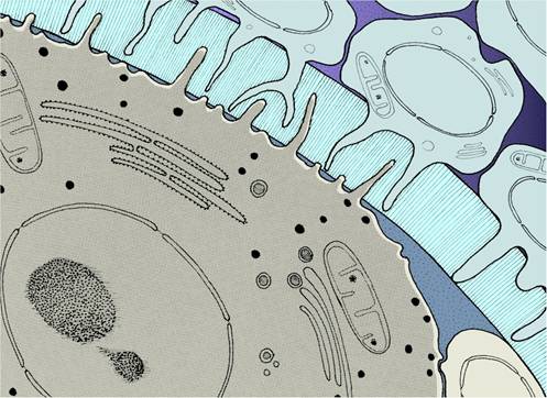
Secondary oocyte
showing at high magnification the relationship between the oocyte
within the zona pellucida and the follicular cells outside the zona. 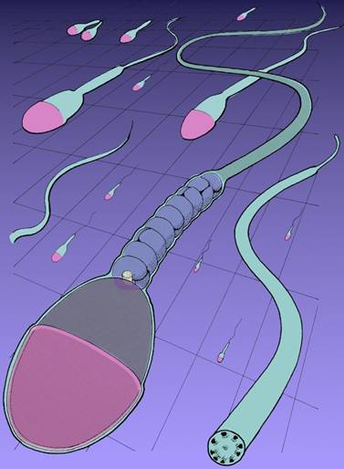
Spermatozoa
showing the head, midpiece, and tail
Sperm and egg viabilityAfter ovulation, the secondary oocyte is viable for up to 24 hours. In the female reproductive tract, sperm are viable for 12-48 hours, although some may persist for 72 hours. Sexual intercourse must therefore take place between 72 hours before and 24 hours after ovulation for fertilization to occur.Sperm movementMillions of sperm are ejaculated but only 1 in 1000 reaches the distal uterine tube. Sperm are motile, and their movement through female reproductive tract is assisted by muscular contractions in the walls of the uterus and uterine tubes. These contractions are stimulated by prostaglandins, present in the seminal fluid.Capacitation of sperm Spermatozoa cannot fertilise the oocyte until they have been capacitated. This process includes removal of a protective layer of cholesterol deposited on sperm plasma membranes by accessory sex glands. The plasma membrane over the head of each capacitated sperm then fuses with the acrosomal membrane. Tiny openings develop at this point, allowing acrosomal enzymes to leak out. The collective activity of the sperm results in dispersal of the corona radiata cells (follicular cells) and penetration through the zona pellucida.
Fertilisation 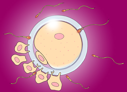
Fertilisation Fertilization is a process that takes several hours. It begins when the fertilising sperm contacts the outer membrane of the secondary oocyte. This triggers recommencement of the 2nd meiotic division of the oocyte. The secondary oocyte divides to form the true ovum and the smaller second polar body. The head of the fertilizing sperm is drawn into the ovum and the genetic material it contains is enclosed in membranes to form the male pronucleus. The nucleus of the ovum becomes the female pronucleus. Fertlisation is completed when the two pronuclei fuse and combine their genetic material to form the genome of the new individual. The fertilised ovum is now called a zygote. In humans, fertilisation occurs in the distal third of the uterine tube (fallopian tube). Spermatozoa may be attracted to the oocyte by chemicals released from it (chemotaxis).Blocking polyspermyTwo mechanisms operate to prevent polyspermy: | fast block - as the 1st sperm cell contacts the oocyte membrane, the membrane depolarises to inhibit further sperm entry | | slow block - when the sperm contacts the oocyte membrane, enzymes are released which harden the zona pellucida, so blocking other sperm from entering |
Nuclear changes in the zygoteAfter fusion of the two pronuclei, a mitotic spindle forms, and the chromosomes of the pronuclei condense and line up on the equator of the spindle.
Cleavage 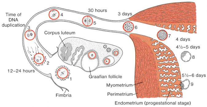
Ovulation, fertilisation, cleavage, implantation – the first week of development The first cleavage division occurs by the end of day 1. Division is by mitosis. The subsequent cleavage divisions occur approximately every 12 hours, giving rise to a ball of cells within the zona pellucida - the morula. MorulaThe morula is nourished by secretions from the epithelium of the uterine tubes. 3 - 4 days after ovulation, the morula enters uterus. The size of morula is constrained by the zona pellucida. Cells in the morula are thought to be totipotent. A cell may be removed for genetic screening at this stage.BlastocystFluid begins to accumulate within the morula. The zona pellucida breaks down and the morula is released. The ball of cells becomes a hllow sphere called the blastocyst. A cluster of cells inside the sphere is called the inner cell mass - it will develop into the embryonic body. The flattened outer cells form the trophoblast, and will contribute to supporting membranes and the placenta. ImplantationA week after fertilization, the blastocyst makes contact with the endometrial lining of the uterus. Most conceptuses implant high on the back wall of the uterus, but sometimes implantation occurs in an ectopic location.Implantation At the site of implantation, the endometrium responds with the decidual reaction. The trophoblast cells release enzymes to assist implantation. Implantation is completed after several days.Multiple conceptionsUsually, during a menstrual cycle only 1 oocyte is released from one of the ovaries. During the next cycle, usually ovulation occurs frm the other ovary. Sometimes, more than one egg is ovulated - if each is fertilised by a separate sperm, this gives rise to non-identical or fraternal twins (or triplets, quadruplets etc). Occasionally, two (or very rarely more) babies originate from a single zygote to form genetically identical twins. Incompletely separated twins are called conjoined twins.Human Chorionic GonadotrophinTrophoblast cells produce a hormone called human Chorionic Gonadotrophin or hCG. hCG enters the maternal circulation and maintains the corpus luteum. This ensures a continuing supply of progesterone and oestrogens which sustain the endometrium. Detection of hCG in the maternal urine forms basis of some home pregnancy test kits. |
|
    
|
