body cavities

Home
Up
cardiovascular system
respiratory system
skeletal system
muscular system
digestive system
urinary system
reproductive system
body cavities
pharynx
face and palate
nervous system
eye and ear

When you have completed this section, you should be able to:

bullet

describe the origin and form of the intra-embryonic coelom

bullet

describe the subdivision of the intra-embryonic coelom into four sealed compartments: 2 pleural cavities, 1 pericardial cavity, and 1 peritoneal cavity.

horizontal rule

 body cavities

There are of course many cavities in the body: nasal cavities, cranial cavity, oral cavity, abdominal cavity - to name just a few. But the term body cavities is generally used to refer to a specific set of cavities, all derived from the intra-embryonic coelom. They are:

bullet

the pleural cavities

bullet

the pericardial cavity

bullet

the peritoneal cavity.

Each of these cavities is extensive in terms of surface area, but in terms of volume amounts to only a few ml. in a healthy person - only a film of fluid separates visceral and parietal layers of the serous membranes bounding these cavities.

intra-embryonic coelom

Recall that the intra-embryonic coelom is a medially-directed cleft that splits the lateral mesoderm into two layers: somatic and splanchnic mesoderm. The intra-embryonic coelom extends forwards, around the front end of the neural plate to form a continuous U-shaped cleft from one side of the embryo to the other. (If you are small enough, you could walk in from one side of embryonic disc, through this curving tunnel, and out at the other side.) The anterior region of the U-shaped coelom is separated from the extra-embryonic coelom by a transverse bar of embryonic mesoderm which will later become the septum transversum. Remember that folding of the embryonic disc will bring this septum to a position caudal to the heart, where it will contribute to development of the liver and diaphragm. This anterior part of the intra-embryonic coelom will become the pericardial cavity, and the heart develops in close relationship with it.

By the time the heart has started to beat, the intra-embryonic coelom has two major compartments, partially separated from each other by the septum transversum: the pericardial cavity and the peritoneal cavity.

pericardio-peritoneal canals

In the mid-embryonic period, the pericardial and peritoneal cavities still communicate with each other by way of two channels that exist behind (dorsal to) the septum transversum. These are the pericardio-peritoneal canals. Later, the lung buds grow down into these canals by passing behind the heart.

In addition, the peritoneal cavity is still in communication with the extra-embryonic coelom that extends along the umbilical cord. This latter communication playes a significant role during development of the midgut - remember the 'physiological herniation' of the midgut loop during weeks 6-12.

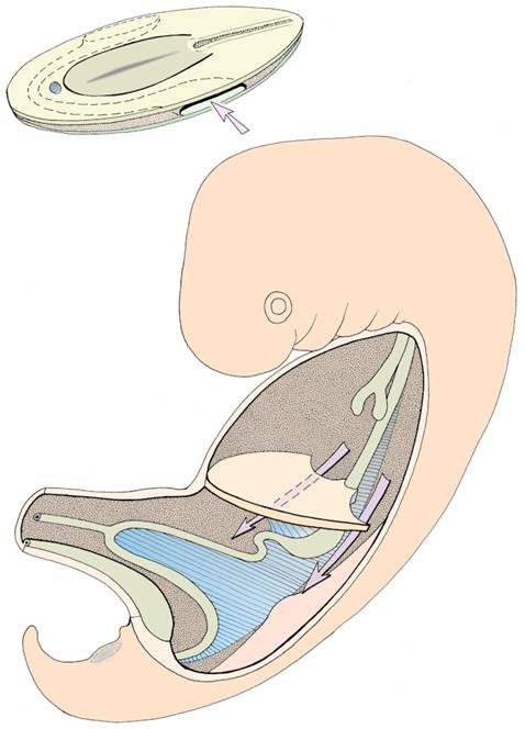
 Intraembryonic coelom - note the septum transversum partially subdividing the thoracic cavity (above) from the abdominal cavity (below). The arrows indicate the pericardio-peritoneal canals.

horizontal rule

further subdivision of the coelom

Final subdivision of the original coelom into pericardial, pleural, and peritoneal portions depends on two events:

bullet

formation of the pleuro-peritoneal folds which close off the canals dorsal to the septum transversum. These folds arise from the mesonephric ridges caudal to the septum, and separate the pericardial cavity from the peritoneal cavity. (A failure in this process can result in congenital diaphragmatic hernia at a latter stage.)

bullet

as the lung buds continue to develop and grow in size, they begin to extend laterally and ventrally until they almost completely surround the heart. At the same time, a membrane is drawn in medially from each side towards the heart. In the free edge of each of these pleuro-pericardial folds lies an anterior cardinal vein, which is draining blood back to the heart from the cranial half of the embryo. These folds separate the pleural cavities from the pericardial cavity.

The continuation of the extra-embryonic coelom into the umbilical cord usually becomes obliterated after return of the midgut to the abdominal cavity, so that the peritoneal cavity also becomes sealed off.

horizontal rule

Questions on development of the body cavities:

  1. What might happen if a baby is born with a large defect in its diaphragm?

  2. Are there any differences in the innervation of visceral and parietal layers of the serous membranes which line the body cavities?

  3. What are the functions of the 4 main body cavities?

  4. How are the different layers of the pericardial sac formed?

horizontal rule

Home Descriptive Contemporary Body systems Birth defects